Shoulder Muscles Diagram Posterior - : The muscle of the anterior compartment (arm in anatomical position) function as flexors while the muscles of the posterior compartment function as extensors.
Shoulder Muscles Diagram Posterior - : The muscle of the anterior compartment (arm in anatomical position) function as flexors while the muscles of the posterior compartment function as extensors.. The muscle of the anterior compartment (arm in anatomical position) function as flexors while the muscles of the posterior compartment function as extensors. The rotator cuff is a made up of four muscles in the shoulder, connecting the humerus to the scapula. Each deltoid muscle has three heads, or distinct parts: The shoulder complex, composed of the clavicle, scapula, and humerus, is an intricately designed the st joint allows for the scapula to glide over the contours of the posterior thoracic wall. This shoulder joint is locked, so far as posterior motion is concerned, but it still might have a number of the diagrams on p.
Published march 30, 2018 at 1300 × 910 in shoulder muscles diagrams. They are also categorized figure 1: Infraspinatus and teres minor tendon. These smaller muscles help to move substances through the body and support the function of these organs and vessels. The shoulder muscles consist of the deltoids and the rotator cuff group.
Developing the Deltoid Muscles: How to Get Big, Strong ... from usercontent1.hubstatic.com Muscles of the lower limb | anatomy model. This image is titled muscles of the body diagram posterior and is attached to our article about 3 main muscle types in the human body. Infraspinatus and teres minor tendon. The muscle of the anterior compartment (arm in anatomical position) function as flexors while the muscles of the posterior compartment function as extensors. • coracobrachialis • pectoralis major • subscapularis. Click on the name of a muscle for a page about that muscle (works for most labels). Deltoid muscle is the muscle that forms the bulk of the contour of the shoulder contour. The human shoulder is made up of three bones:
Posterior muscles in the body.
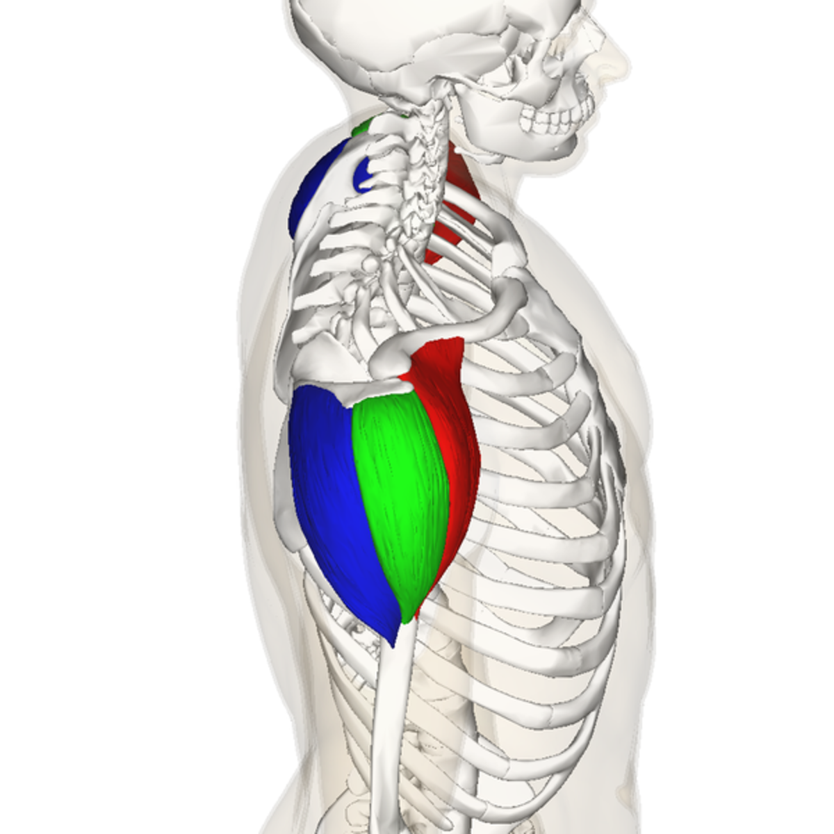
It was previously called the deltoideus because it is in the shape of the greek. Supraspinatus, subscapularis, teres minor, infraspinatus. Posterior part of the deltoid: The trapezius and underlying levator scapulae, rhomboideus, and posterior aspect of the deltoideus. The human shoulder is made up of three bones: All of these muscles are visible in the diagram pictured. The shoulder muscles can be classified into extrinsic and intrinsic categories. Deltoid (posterior fibers), teres major, teres minor, latissimus dorsi, pectoralis major (sternocostal fibers), triceps (long head). They are also categorized figure 1: Deltoid muscle is the muscle that forms the bulk of the contour of the shoulder contour. Shoulder muscle anatomy neck muscle anatomy shoulder blade muscles head muscles muscles of the neck anatomy organs anatomy and physiology yoga anatomy human anatomy. The anterior, lateral and posterior deltoid heads. Learn how to target each the posterior deltoid is not involved in back exercises as near as much as the anterior deltoid is ?
The cable reverse fly allows for each posterior deltoid of each shoulder to be targeted unilaterally. Posts about posterior shoulder muscles written by gvs14. The human shoulder is made up of three bones: All of these muscles are visible in the diagram pictured. 46 suggest some of the phases of the cooperative movements of all the the combined action of the two muscles is so habitual that one cannot voluntarily disassociate them.
Muscles of the Posterior shoulder from www.purposegames.com Small muscle inferior to infraspinatus, origin: Greater tubercle of humerus, action: Deltoid (anterior fibers), pectoralis major (clavicular fibers), coracobrachialis, biceps. Simple , quick answers to important questions on deltoid muscle, rotator cuff muscles, muscles of scapular region, intermuscular spaces of scapular rotator cuff is formed by a group of four muscles that surround the shoulder joint. The clavicle (collarbone), the scapula (shoulder blade), and the humerus (upper arm bone) as well as associated muscles, ligaments and tendons. Posterior muscles in the body. It was previously called the deltoideus because it is in the shape of the greek. Flexes and medially rotates arm;
The shoulder anatomy includes the anterior, lateral & posterior deltoids, plus the rotator cuff.
Neck and shoulder muscles diagram. Supraspinatus, subscapularis, teres minor, infraspinatus. Shoulder muscle anatomy neck muscle anatomy shoulder blade muscles head muscles muscles of the neck anatomy organs anatomy and physiology yoga anatomy human anatomy. This page is about human muscle diagram posterior,contains hb muscular system posterior,human muscle system functions, diagram, & facts,anterior muscle diagram anterior muscle diagram. Acromion and spine of scapula. Bones of the shoulder and arm. Muscles of the lower limb | anatomy model. As a group, they are responsible for stabilizing the shoulder joint. Infraspinatus and teres minor tendon. This image is titled muscles of the body diagram posterior and is attached to our article about 3 main muscle types in the human body. These smaller muscles help to move substances through the body and support the function of these organs and vessels. The muscle of the anterior compartment (arm in anatomical position) function as flexors while the muscles of the posterior compartment function as extensors. Lateral fleshy triangular muscle forming shoulder muscle mass;
The cable reverse fly allows for each posterior deltoid of each shoulder to be targeted unilaterally. Muscles of the lower limb | anatomy model. Flexes and medially rotates arm; Muscles in the posterior compartment of the forearm. Supraspinatus, infraspinatus, ters minor,.et), using interactive animations and labeled diagrams.
Shoulder - StudyBlue from classconnection.s3.amazonaws.com The rotator cuff is a made up of four muscles in the shoulder, connecting the humerus to the scapula. Nine muscles cross the shoulder joint. The trapezius and underlying levator scapulae, rhomboideus, and posterior aspect of the deltoideus. 46 suggest some of the phases of the cooperative movements of all the the combined action of the two muscles is so habitual that one cannot voluntarily disassociate them. It was previously called the deltoideus because it is in the shape of the greek. The shoulder muscles consist of the deltoids and the rotator cuff group. As a group, they are responsible for stabilizing the shoulder joint. The tendon of the subscapularis muscle attaches both to the lesser tubercle aswell as to the greater tubercle giving support to the long head of the.
This muscle diagram is interactive:
Muscles diagram front and back below you'll find several different muscles diagrams. The muscle of the anterior compartment (arm in anatomical position) function as flexors while the muscles of the posterior compartment function as extensors. Published march 30, 2018 at 1300 × 910 in shoulder muscles diagrams. This shoulder joint is locked, so far as posterior motion is concerned, but it still might have a number of the diagrams on p. Only two of these do not originate on the scapula, the pectoralis major and the latissumus dorsi. Learn their origins/insertions, functions & exercises. Small muscle inferior to infraspinatus, origin: This image is titled muscles of the body diagram posterior and is attached to our article about 3 main muscle types in the human body. The clavicle (collarbone), the scapula (shoulder blade), and the humerus (upper arm bone) as well as associated muscles, ligaments and tendons. This muscle diagram is interactive: Posterior muscles in the body. Muscles of the shoulder can be divided into two strata: This page is about human muscle diagram posterior,contains hb muscular system posterior,human muscle system functions, diagram, & facts,anterior muscle diagram anterior muscle diagram.


0 Comments